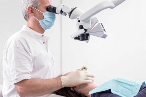
M320 D
Dental microsurgery involves the use of magnification and illumination for an enhanced visualization of anatomical details and fine structures. The dental microscope is an essential instrument used in dental procedures and dental surgery, allowing optimal assessment, preservation and maintenance of tooth structure and oral tissue, as well as minimally invasive procedures.
Leica Microsystems offers cutting-edge Dental Surgical Microscopy solutions, combining world-renowned optics, optimal ergonomics and maneuverability, and ease of use to support dental professionals in achieving surgical excellence.


Applications of Dental Surgical Microscopy
The use of microscopes in dental surgery has a wide range of applications, since it allows an enhanced visualization of fine anatomical details and tissue structures. By using higher magnification and seeing more, dental professionals can apply their skills more precisely and optimize their workflow. Some of the most common applications of Dental Surgical Microscopy, also referred to as micro-dentistry, include the following:
- Endodontics and endodontic surgery
- Periodontics and periodontal surgery
- Implantology
- Prosthodontics and aesthetic dentistry
- Restorative dentistry
- Oral surgery
- Routine dental techniques
Challenges of Dental Microsurgery
A dental microscope should provide optimal light intensity and depth of field, while achieving sufficient resolution when working in deep or narrow cavities, such as during root canal treatment.
In micro-dentistry procedures, it is essential to achieve optimal and precise control of dental instruments under high magnification to avoid damaging the dentine walls or other tissues.
Moreover, it is important to achieve visualization of anatomical details in vivid colors to ensure their correct differentiation, such as during removal of pathologic tissue during dental procedures or surgeries.
Maintaining a natural and comfortable working posture is another major challenge in dental microsurgery, as poor ergonomics can lead to significant stress and fatigue during lengthy procedures.
M320 Dental Microscope
With Integrated 4K Camera
Enjoy better visualization during surgical and non surgical dental procedures, benefit from increased physical comfort, and show your expertise to peers and patients with astounding 4K images.
Inspired by dentists ‐ With the M320 dental microscope you can achieve better treatment results and optimize outcomes through high precision microdentistry. To help you share your expertise with patients and peers you can record your procedures in impressive 4K image and video quality.
Developed for dentists - The ergonomic microscope design of the M320 enhances your physical comfort and thus increases efficiency in different fields including restorative dentistry, endodontics, prosthodontics and aesthetic dentistry as well as periodontics and implantology.
Not all products or services are approved or offered in every market and approved labeling and instructions may vary between countries. Please contact your local Leica representative for details.
Reveal details in cavities
With the M320 dental microscope you can see just what you must see in minute anatomical detail. Even in deep or narrow cavities, you can apply your skills precisely and efficiently.
- Differentiate anatomical detail easily thanks to the powerful LED illumination with high color perception and reproduction
- Benefit from a bright view in endodontic applications - two LED paths provide a homogeneous illumination
- Choose from a complete range of objective lenses including the M320 MultiFoc Objective
- Change magnification smoothly and thus minimize workflow interruption thanks to the optional 5-step magnification changer
- Enjoy and share brilliant images and videos thanks to the fully integrated 4K camera
Strengthen patient trust
Reinforce trust by walking your patient and their family through the steps of a procedure using live images or video playback.
Let the M320 with integrated 4K camera support you to show them they are in good hands.
With its variety of viewing options, the M320 facilitates patient communication.
You can involve your patients, illustrate different treatment options to them, and make them part of the decision process.
Ultra-high resolution images can be easily transferred and archived in the patients’ files for recall at any time.
Enhance your comfort
You can adapt the M320 to your body frame and preferences, positioning it quickly and accurately with the lightest touch.
This reduces the potential strain of a hunched working position and harsh movements, so that you can diagnose and treat your patients without distractions.
The M320 helps you to focus entirely on your patient, during surgical procedures as well as in non-surgical treatments.
- Two binocular tubes: The 45˚ one for a comfortable view in a fixed, standard position, and the 180˚ one for more comfort and flexibility with more viewing angles.
- Two dedicated ergonomic accessories: ErgoWedge providing a tiltable viewing position and the ErgonOptic Dent to extend the reach of the microscope and swivel the optics carrier to any angle.

Easy viewing, recording and sharing with the Leica View App for the M320 dental microscope
- Stream the live microscope view from the M320 dental microscope to a mobile device
- Capture images and video directly from the mobile device
- Access recorded pictures and videos from the SD-card gallery of the M320
- Download assets to a mobile device to have them conveniently at hand for image-supported patient communication
- The Leica View App is available free of charge. For iOS devices you can download it from the App Store and for Android from Google play.
Only with optionally ordered WiFi-dongle. Streaming resolution to todays’ generations of mobile devices: 720p30


