
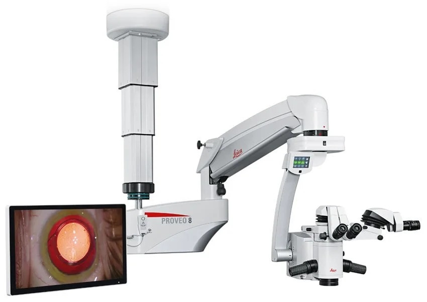
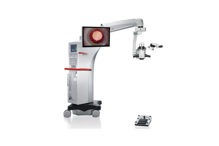
Proveo 8
Leica Microsystems offers a range of state-of-the-art ophthalmology microscopy solutions that support ophthalmic surgeons in achieving optimal patient outcomes in anterior and posterior segment surgery. Our ophthalmology microscopes are world-renowned for premium optics and illumination that provide outstanding image quality. Designed to ensure a seamless workflow and support ergonomics in the OR, they optimally respond to the surgeon’s needs throughout each step of the procedure.
Leica ophthalmology microscopes form the platform for complete ophthalmology imaging and workflow solutions by seamlessly integrating advanced imaging technologies, such as Optical Coherence Tomography (OCT) and high-definition display and documentation features.


Applications of Ophthalmic surgical microscopes
Ophthalmic surgical microscopes reveal finest anatomical details and facilitate surgical maneuvers during anterior and posterior segment surgery. Their use in ophthalmology has a wide range of applications, from diagnostics to complex surgical procedures. Some of the common applications of ophthalmic microscopes in surgery include the following:
Anterior Segment Surgery
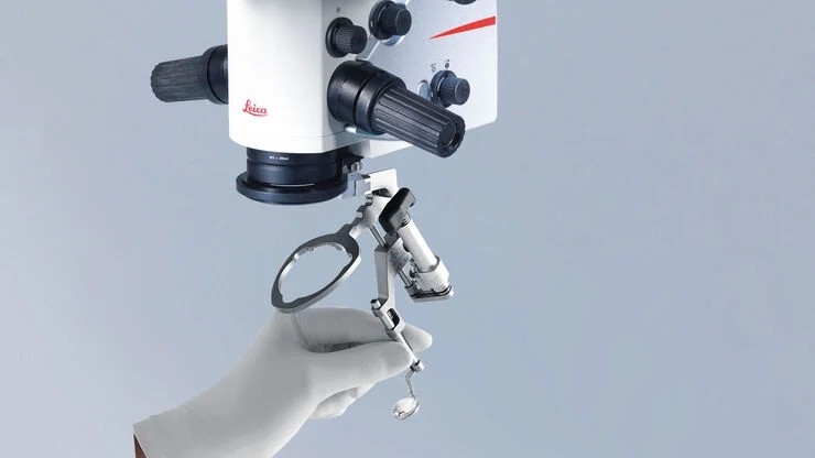
- Cataract surgery
- Refractive surgery
- Glaucoma surgery
- Corneal surgery
- Laser eye surgery
Posterior Segment Surgery (Vitreoretinal Surgery)
- Vitrectomy
- Retinal detachment repair
- Macular hole repair
- Maculopathy surgery
- Posterior sclerotomy
- Radial optic neurotomy
- Macular translocation surgery
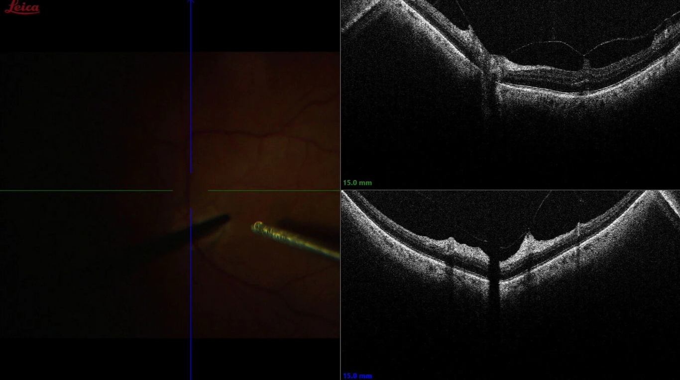
In addition, OCT solutions can be used for pre-operative assessment, as well as for intraoperative visualization and confirmation of tissue response to surgical maneuvers.

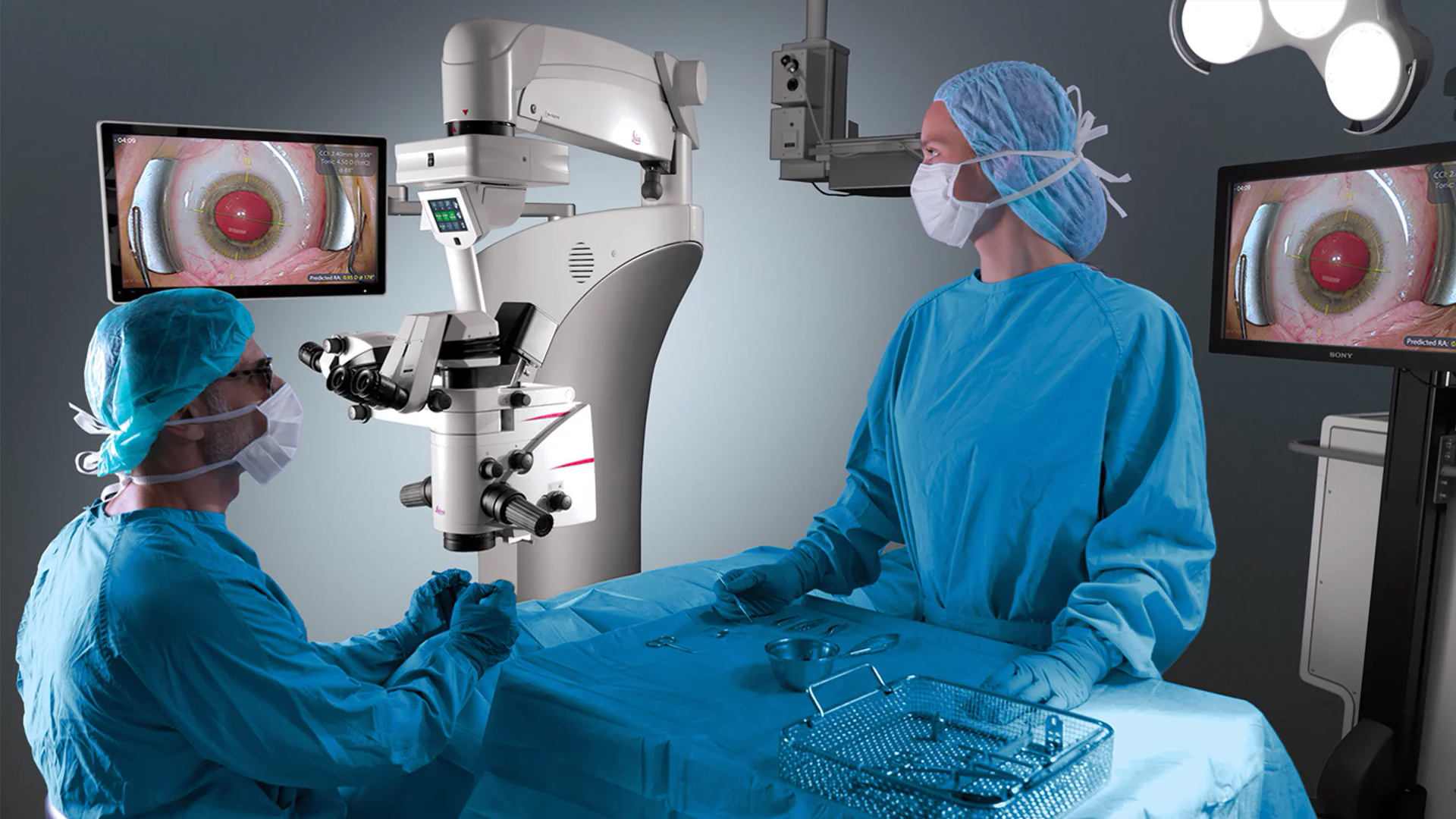
Stable Red Reflex, Consistent Images
Focus on your cataract surgery and be confident that you’ll have an constant, uncompromised view.
You can rely on stable red reflex and optimal image contrast thanks to CoAx 4, coaxial LED illumination.
Plus, all observers – surgeon, assistant, and camera – work with the same enhanced view during the entire surgical procedure.
- Four individual coaxial beam paths and linked zoom system, deliver straight red reflex for all observers.
- Adjust the illumination diameter to each individual patient’s eye – so no light is lost. This means lower light can be used while still achieving maximum contrast.

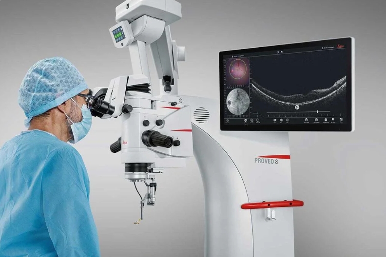
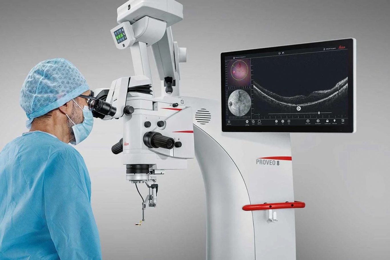
Focus on Perfection: Proveo 8 with built-in intraoperative EnFocus Optical Coherence Tomography (OCT)
Apply your skills with even greater confidence during complex anterior and posterior segment surgeries. EnFocus intraoperative OCT provides you:
- Greater insight with bright, sharp images of previously hidden subsurface tissue details.
- Immediate confirmation in real-time so you can see how ocular tissue is reacting to your surgical maneuvers.
- Maximum freedom, because OCT is now fully integrated into your Proveo8 and into your workflow
More detail, less refocusing
See more detail in one view and reduce time-consuming refocusing thanks to groundbreaking FusionOptics technology from Leica Microsystems.
FusionOptics technology overcomes previous optical limitations by combining high resolution and depth of field. You benefit from one crisp, texture-rich image from the edge of a detached membrane to the retina.
The Technology of FusionOptics
- Two separate beam paths
- One beam path provides 40% increased depth of field
- The other beam path provides high resolution
- The brain merges the two images to a single optimal spatial image